Optic Nerve excavation

Lima, Peru

Prof Mario de La Torre

Graduated as an ophthalmologist 32 years ago, in 1993 he obtained his Degree of Doctor of Medicine at the Julius Maximilians University of Würzburg Germany, with a mention in Ocular and Orbital Ultrasound, obtaining the Magna cum Laude qualification.

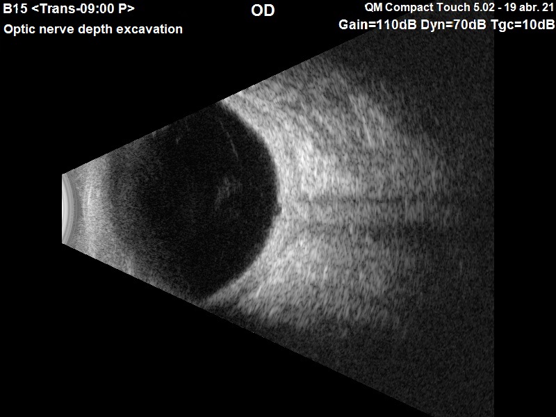
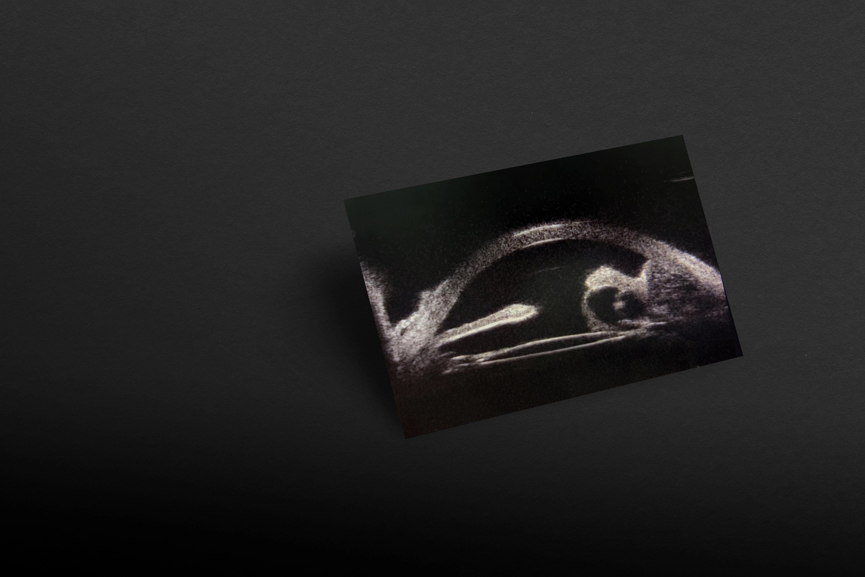
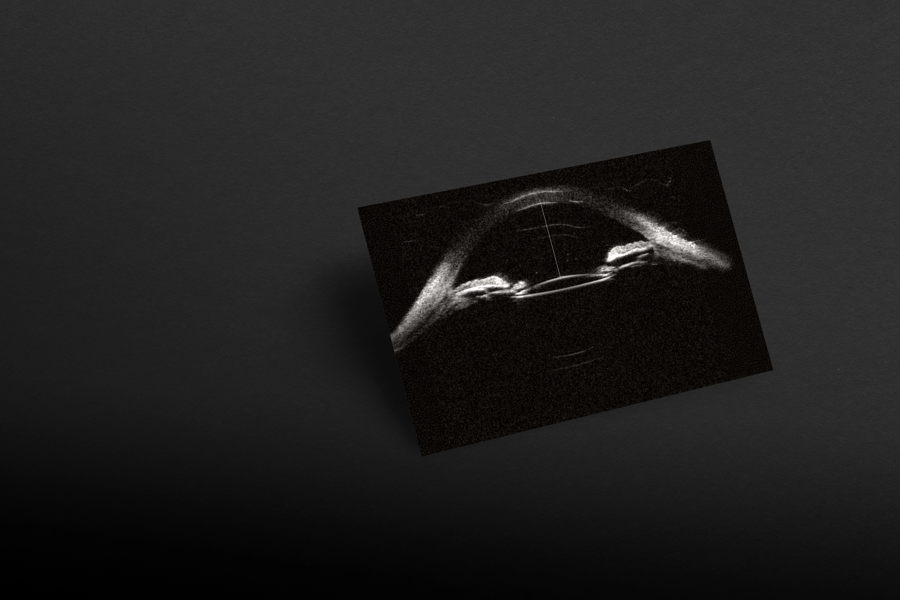
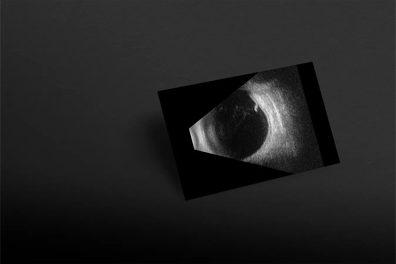
In routine examinations prior to cataract surgery, it is important to determine the state of the macula and the optic nerve to rule out pathologies that may worsen the patient's visual prognosis after surgery.

#1

Ask for the right diagnosis

<p>Ask for the right diagnosis</p>

Despite the fact that ultrasound is not the diagnostic method of choice for glaucoma, the fact of observing marked and very especially asymmetrical excavations, can lead us to be more careful and guide our efforts to determine if it is a glaucoma excavation or a mega papilla through other studies.

In this case, the examination with the Compact Touch® allows us to observe the marked papillary excavation of both eyes, which at the same time is symmetrical. This type of examination is important as a complement to the study of the patient and alerts us to possible pathologies, in this case a more in-depth study must be done.

No content found

Interested by range ?