Anterior segment

Anterior Segment Tumor

Lima, Peru

quezada 675c486fea9fb352432771

Dr Gabriela Quezada G.

DLT Centro de Diagnostico Oftalmologico - Lima, Peru

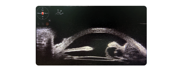
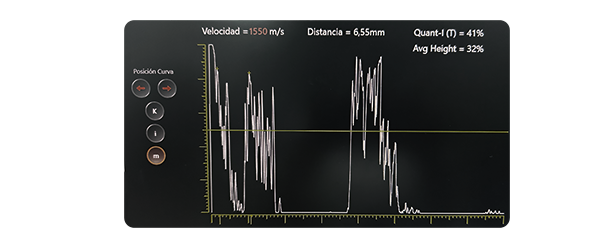
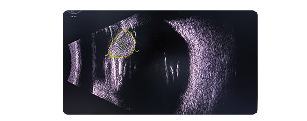
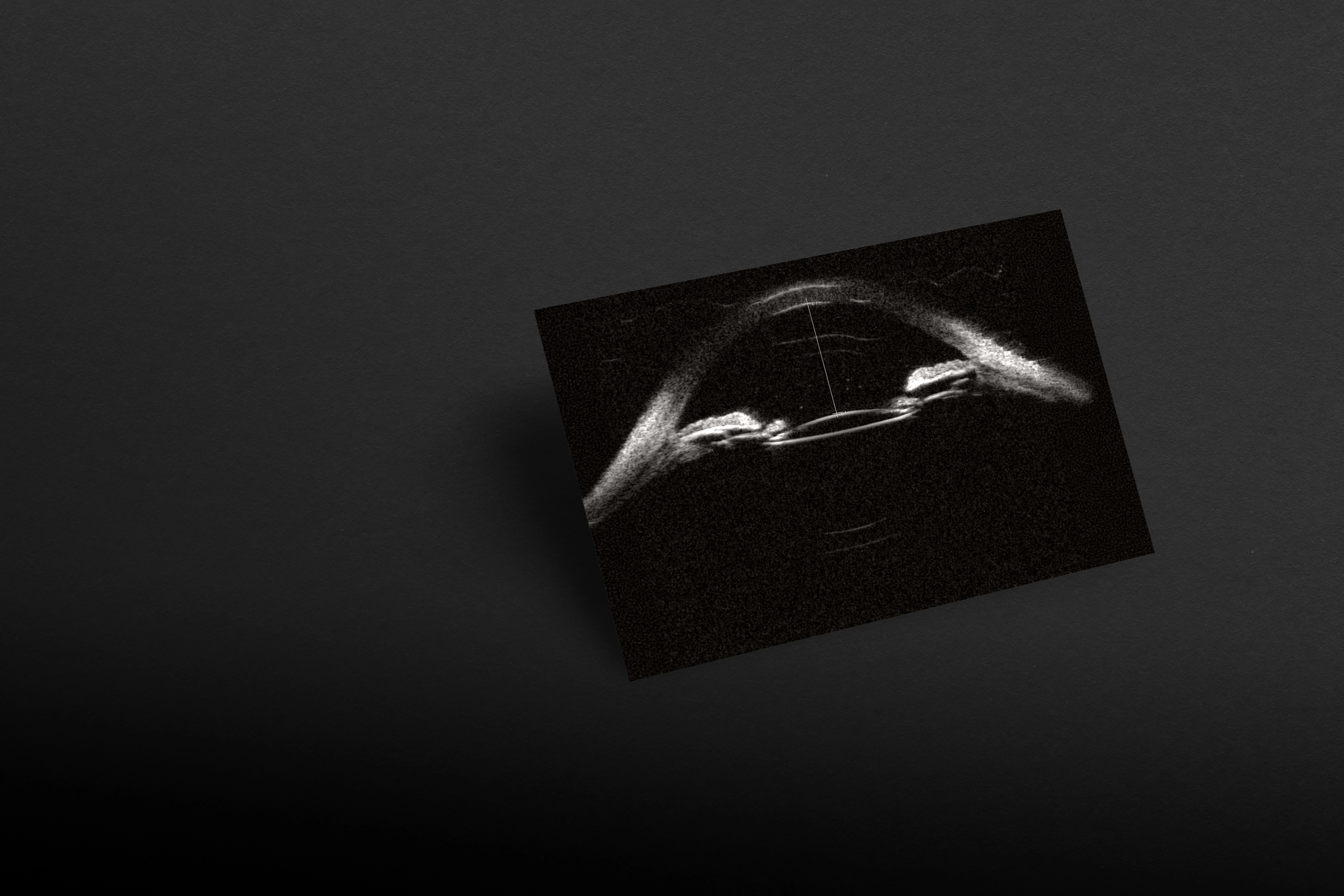
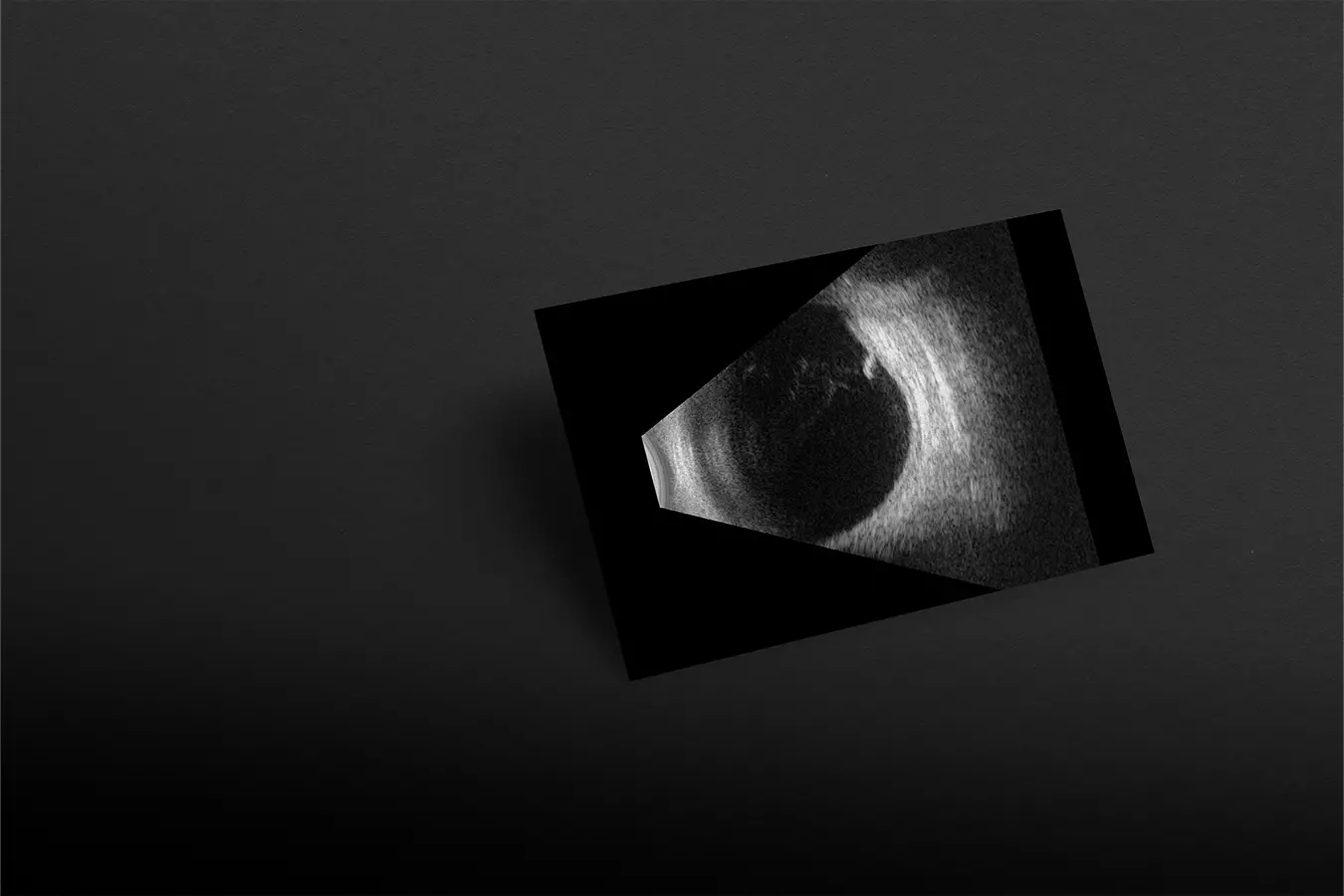
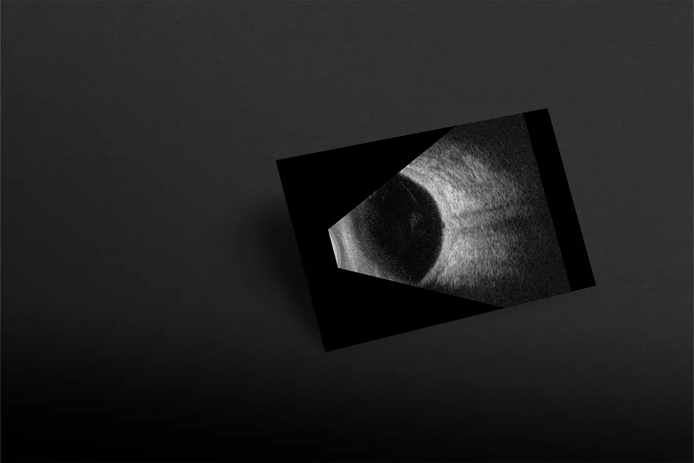
A 66-year-old male patient presents with ocular pain and a reduction in visual acuity to 20/50 in the right eye (OD), one week after undergoing cataract surgery in the left eye (OS). Clinical examination reveals an elevated intraocular pressure (IOP) of 32 mmHg in OD. Slit-lamp evaluation shows conjunctival chemosis, the presence of anterior chamber cellularity, and anterior synechiae leading to angle closure over 3 clock hours. These findings raise concern for postoperative inflammatory angle closure.

#1

UBM

ubm scan

A tumor of the ciliary body was identified, accompanied by multiple iris cysts causing anterior displacement of the iris root, leading to angle closure and contact with the intraocular lens (IOL).

#2

Standardized A mode

standardized a mode scan

Ultrasound A-mode confirmed the presence of a melanoma in the ciliary body, characterized by low internal reflectivity and vascularity — features typically observed in this type of tumor.

#3

B mode

b mode scan

The lesion was identified using B-mode ultrasound based on its reflectivity, which also allows for monitoring tumor growth over time.

#4

Conclusion

UBM, combined with B-mode and standardized A-mode ultrasound, can differentiate between benign and malignant lesions based on reflectivity patterns.
 

The ability to visualize and measure tumors using UBM allows for better surgical planning and more accurate prognosis. It can also be used to track tumor growth over time, ensuring that any changes in size or structure are addressed in a timely manner.

Get this case study 
in PDF format: here