Ocular surface

Vernal keratoconjunctivitis (VKC) in children

Saint-Étienne, France

dr trone 6499a7a20655e629300810

Dr. Marie-Caroline Trone

Hospital-based Ophthalmologist at Saint-Étienne University Hospital – France

Diagnosis and treatment of a child with vernal keratoconjunctivitis (VKC) associated with meibomian gland dysfunction (MGD). A full diagnosis was made via clinical examination and meibography using C.Diag®. Intense Pulsed Light treatment using the C.Stim® IPL system was started for the patient. After 6 months, meibum quality had improved with an expressible, fluid consistency.

Anamnesis :

Paul, an 8-year-old schoolboy, sought urgent medical attention after experiencing red, painful eyes for several days (in April). He was highly sensitive to light. He was in good health and not taking any medication but has seasonal pollen allergies.
#1

Clinical examination

The ophthalmological examination showed the following results:

OD


OS

+ 1.50

Refraction

+ 1.25

Not possible

VA

Not possible

Not possible

IOP

Not possible

The child was extremely photophobic and struggled to open his eyes. The slit-lamp examination was difficult.


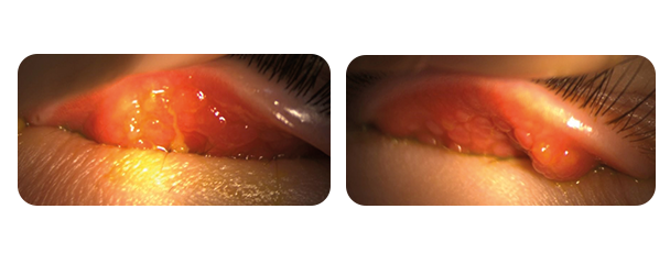
Giant papillae were, however, visible during eyelid eversion. The rest of the examination revealed a clear cornea in both eyes with no vernal plaque or Trantas dots. A diagnosis of vernal keratoconjunctivitis (VKC) was made.

#2

Initial treatment

Initial treatment involved a short course of rapidly tapering high-dose topical corticosteroids, eye bathing with saline solution several times a day, ZALERG® topical antihistamine and mast cell anti-degranulation treatment, along with photoprotection including cap and sunglasses.

The patient was monitored for 48 hours, during which symptom improvement and cortico-dependence were observed.

VERKAZIA® cyclosporine-based eye drops at a dose of 1 drop 4 times a day were introduced.

Allergy testing confirmed a grass allergy in particular and desensitisation treatment was commenced.

Follow-up

Attacks were less frequent but Paul sometimes experienced stinging or burning eyes. Three episodes of chalazion occurred in 2 months.

A further ophthalmological examination was carried out to check refraction under cycloplegia. Minor optical correction of hypermetropia (OD +1.25 and OS +1.00) was prescribed.

#3

Paraclinical examination

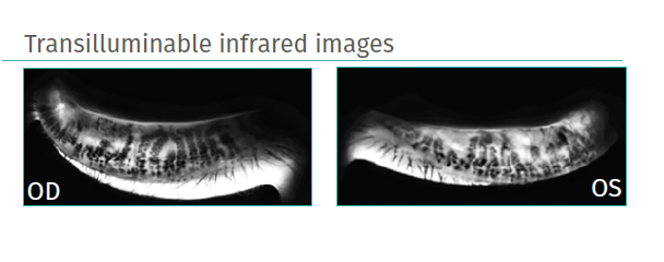
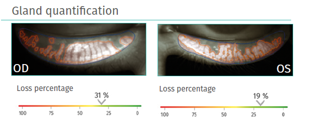
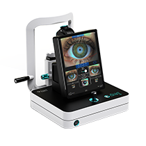
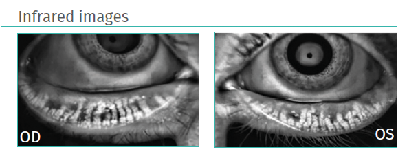
Meibography using C.DIAG was carried out to obtain infrared and transilluminable infrared images. Meibomian gland loss relating to chalazia was observed.


Minor meibomian gland dysfunction was diagnosed with very thick, difficult-to-express meibum.

Follow-up

Eyelid care was difficult to implement, with poor compliance. The meibum was still thick with numerous episodes of chalazion at 6 months.

IPL sessions were offered along with continued eyelid care (with an easier-to-use electrical heated mask).

#4

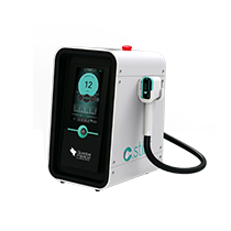
C.STIM IPL treatment in children 

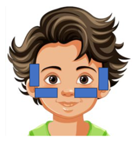
IPL in children involves 3 sessions of just 2 flashes per side. Forceps extraction is not systematically used in young people as it can be perceived as "traumatic".


Follow-up

6 months later, a single episode of chalazion had occurred and there were no episodes of vernal keratoconjunctivitis. Improved eyelid care compliance was observed, as well as improved meibum quality with a fluid and expressible consistency.

Conclusion

It is easy to take meibography images of children using the C.Diag® system.

IPL sessions can be carried out on children presenting with MGD associated with VKC: 

  • Fewer shots on small faces (1 on the temple and 1 on the cheekbone)
  • No forceps extraction

Interested by range Okulære overflatesykdommer?