Cystoid macula edema

Lima, Peru

Prof Mario de La Torre

Graduated as an ophthalmologist 32 years ago, in 1993 he obtained his Degree of Doctor of Medicine at the Julius Maximilians University of Würzburg Germany, with a mention in Ocular and Orbital Ultrasound, obtaining the Magna cum Laude qualification.

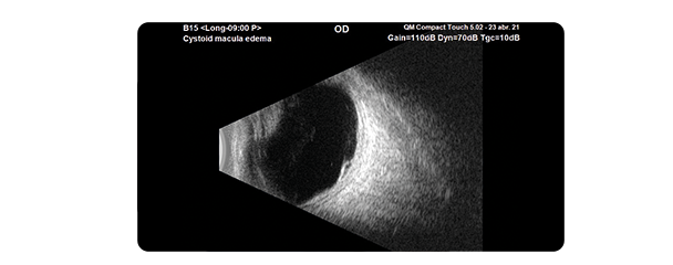
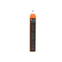
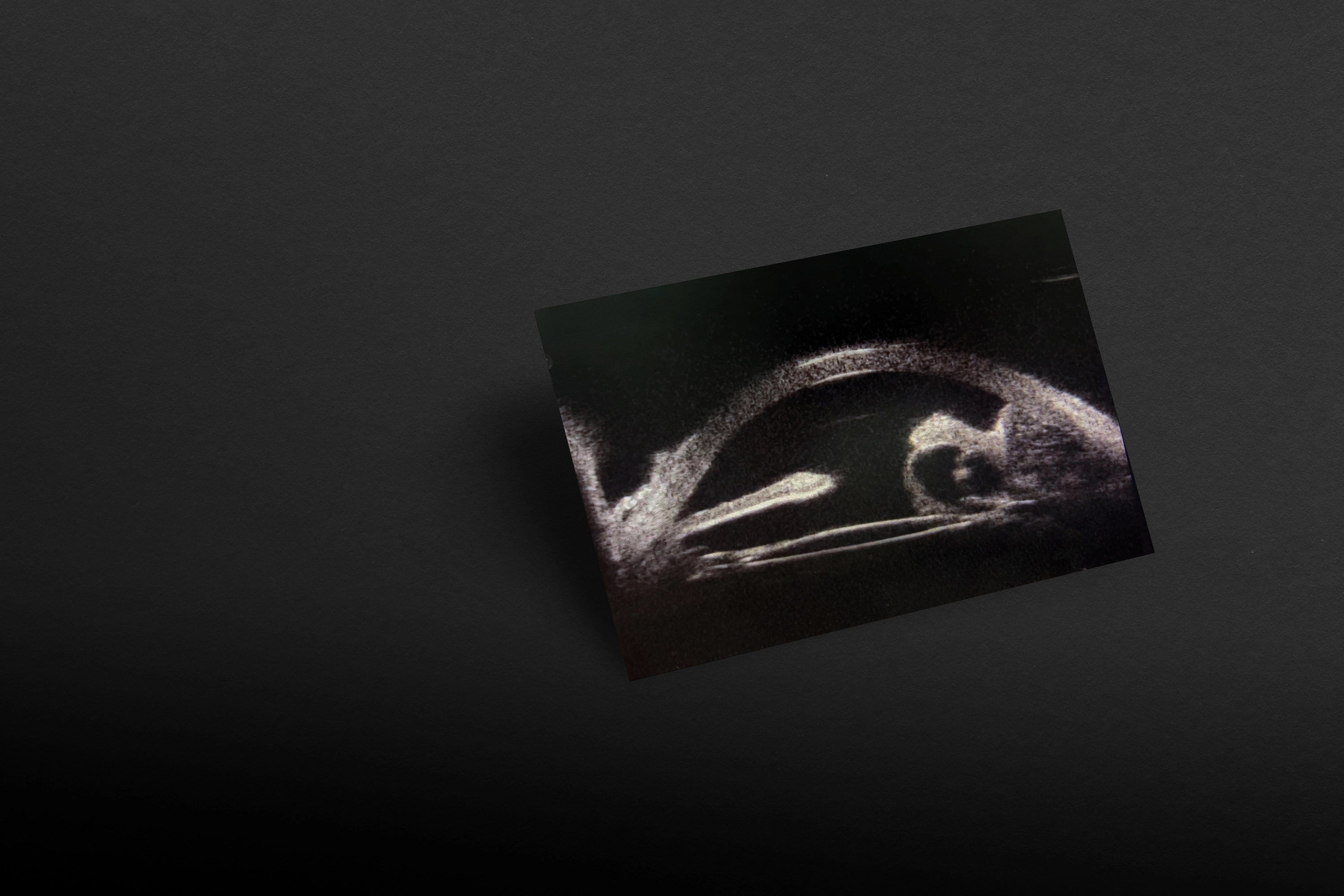
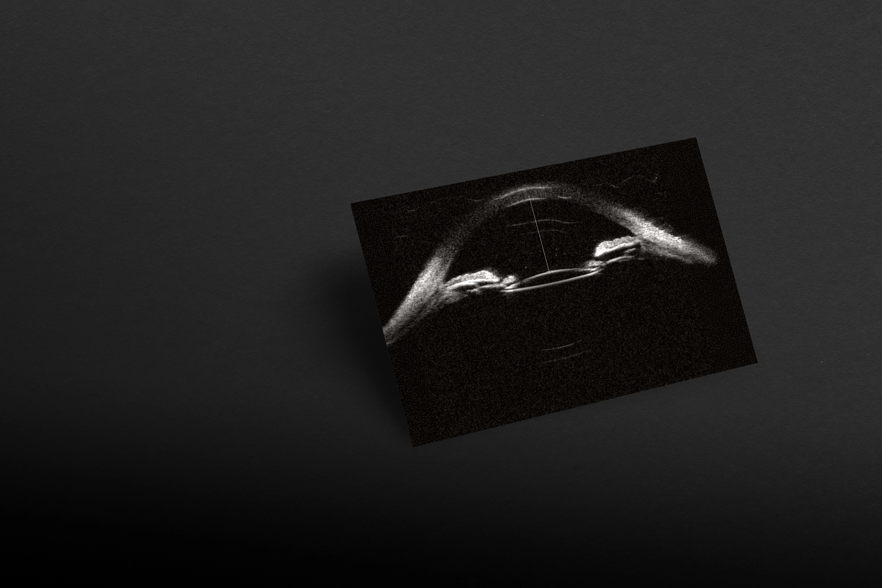
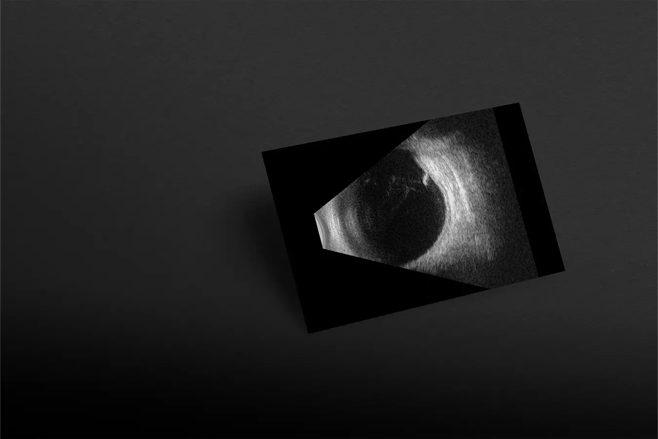
OCT is commonly used to study macular pathology, but what happens if the media are opaque and even with the swept source OCT we cannot see the back of the eye clearly?

#1

Ask for the right diagnosis

<p>Ask for the right diagnosis</p>

In these and many more cases, the ultrasound of the macula, in this case with the Compact Touch®, has the advantage of being able to see “beyond the obvious” by giving the ophthalmologist an examination tool, which has no optical limits and can give an alternative in the study of macular lesions within the ultrasound resolution.

Here we show an elevation of the macular area with well differentiated limits and with small cystic spaces inside, which are visible when zoomed in. An advantage of examining with the Compact Touch®.

No content found

Interested by range ?