Anterior segment

UBM Diagnosis of Phacomorphic Glaucoma

Lima, Peru

Dr Gabriela Quezada G.

DLT Centro de Diagnostico Oftalmologico - Lima, Peru

A 65-year-old male patient presents with pain in the left eye (OS) and a marked reduction in visual acuity, measured at 20/100. Clinical examination reveals an elevated intraocular pressure (IOP) of 30 mmHg. Biomicroscopy identifies an intumescent lens, a narrow anterior chamber, and the presence of iris bombe. A pupillary block is suspected. Gonioscopy confirms 360-degree angle closure.
#1

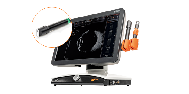
Ultrasound Examination: UBM probe with ABSolu®

<p>Ultrasound Examination: UBM probe with ABSolu®</p>

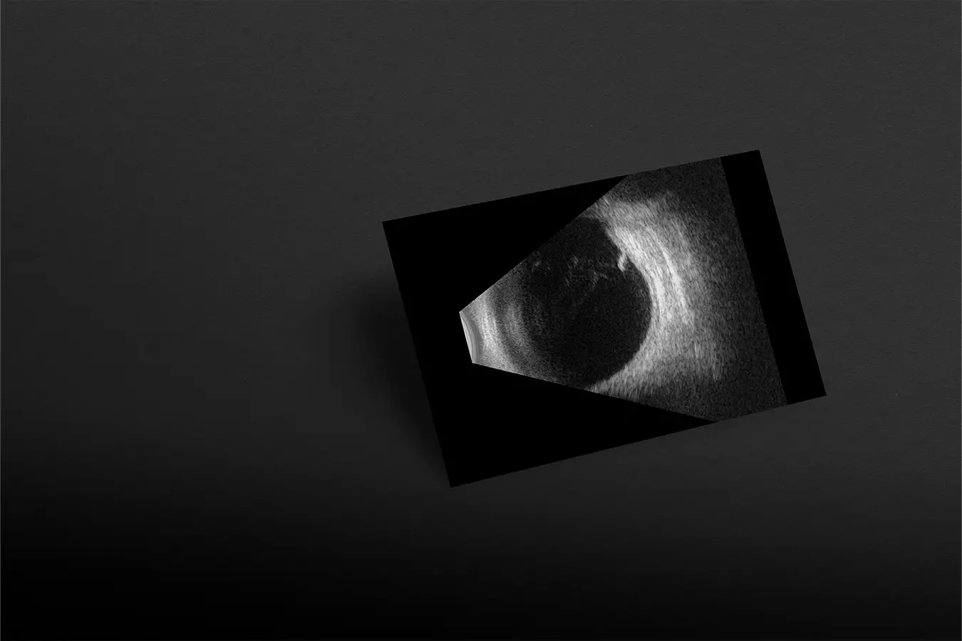
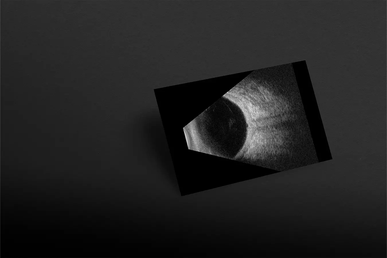
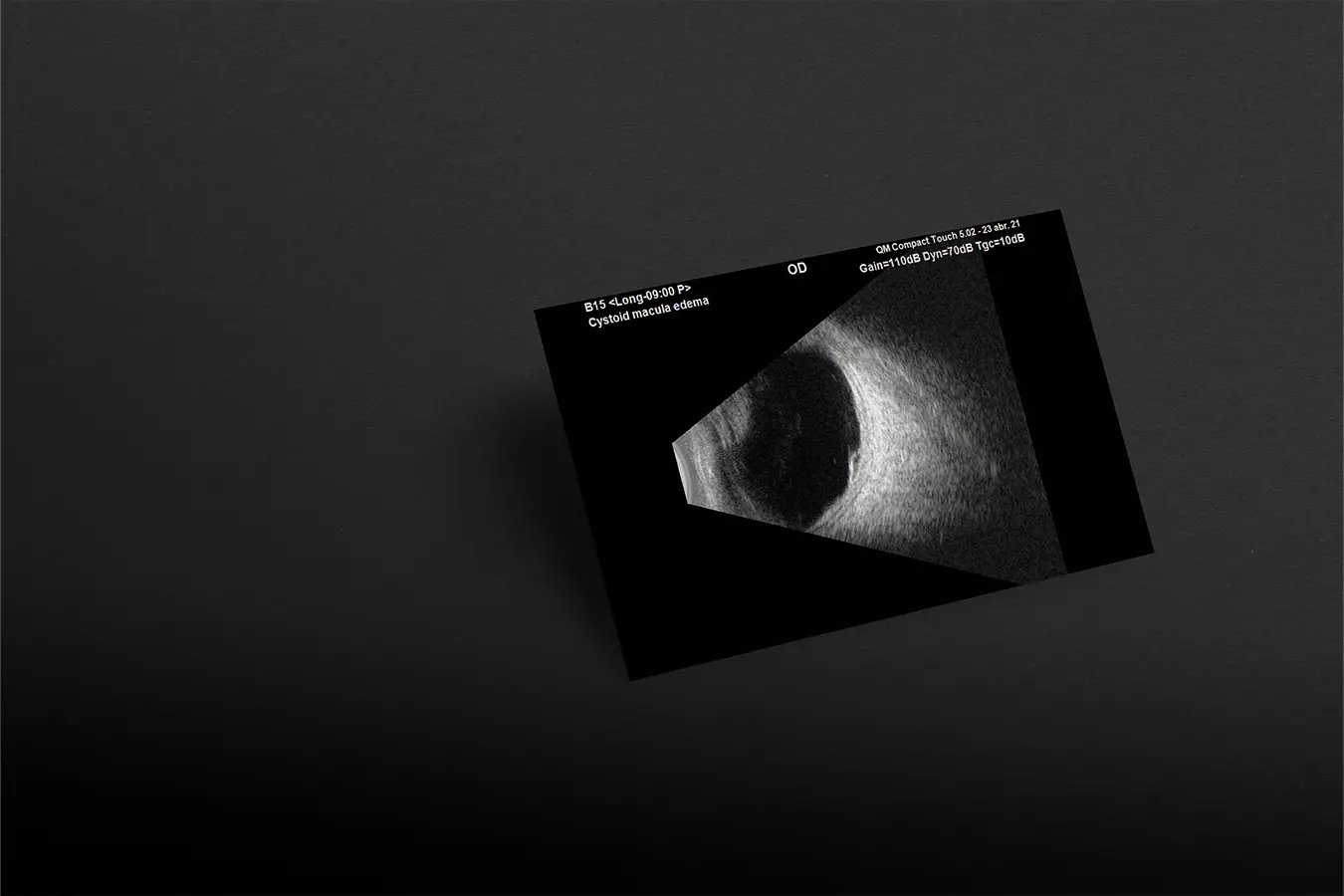
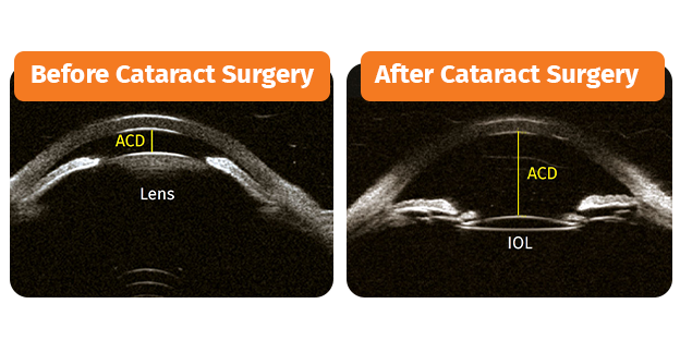
Before cataract surgery, the UBM exams showed the phacomorphic component with pupilar block and angular closure in360 degrees associated to ocular hypertension.

After cataract surgery, the UBM allowed to see the resolution of angular closure and pupillary block, the great depth of the anterior chamber, the identification of centered IOL in the capsular bag and decrease of IOP.

#2

UBM's  Added Value

<p><span style="color:hsl(0,0%,100%);">UBM's &nbsp;Added Value</span></p>

Ocular hypertension and glaucoma are becoming more common in patients with cataracts, in which the increase in lens volume causes the structures to move anteriorly, generating pupillary block and angle closure.

UBM becomes an essential diagnostic tool for the evaluation of structure in real time and for determining the origin of the ocular pathology. In this case of phacomorphic glaucoma, UBM exams value is demonstrated in both preoperative planning and postoperative follow-up of a cataract surgery.


Get this case study 
in PDF format: here

Interested by range Ultraääni?